1) ROM
It is important to regain full ROM by 4-6 weeks post-operatively. Most protocols agree with this. In some cases, ROM may be limited early depending on physician's protocol. But it is important to establish full motion especially into extension by 4-6 weeks post-operatively. ROM exercise may include heel prop, heel slide, wall slide, etc. Stretching calf and hamstring muscles will help to regain knee extension.
2) Quadriceps Straightening
Quadriceps strengthening can be initiated as soon as inflammation is controlled. Initially quad sets with or without NMES(neuromuscular electrical stimulation) is a good way to start firing quad muscle and it can be progressed to straight leg raises (only if knee extension is maintained during exercise). Strengthening exercises for other muscles can also be initiated. It is important to follow physician's protocol and to protect reconstructed graft.
3) Gait
Normal gait should be established as the injured athlete can tolerate. If he/she can't walk normally, they should be placed on the crutches. Weight shift exercise and gait training on treadmill can help to re-establish normal gait as well as walking in a pool.
4) Strengthening (closed kinetic chain)/balance exercise
CKC strengthening exercise can be initiated later in this phase of rehab. It includes partial squat, wall squats, leg press, single leg balance, TKE, etc. Again each physician has their own protocol, so it is important to follow it.
The goals of this phase of rehab will be 1) regain full ROM, 2) establish normal gait, 3) start strengthening exercises that include CKC exercise and balance training. Also, strengthening training should be pain-free and inflammation should be under control.
Wednesday, May 25, 2011
Monday, May 16, 2011
Rehabilitation after ACL Tear: Immediately Post-Op
Immediately after surgery, the knee will be swollen and inflamed. For the first 10 - 14 days after surgery, the main focus is on reducing the inflammation. Depending on the surgeon, formal rehabilitation will be held for the first 2 weeks. The injured athlete should be careful with the woulds from the portal even though they might not be large. There is still a risk of surface infection at the portal sites. The knee might be placed in a rigid knee brace to protect the reconstructed knee. Or the athlete may not even wear any type of brace depending on the surgeon's preference. CPM (continuous passive motion) and PROM (passive range of motion) exercise can be initiated immediately after surgery. However, it is dependent on surgeon's protocol. Ice and NSAIDs are used to reduce and control inflammation. Elevating the knee above the heart may also help to reduce swelling.
Friday, May 13, 2011
Rehabilitation after ACL Tear: Pre-Operative
Anterior cruciate ligament (ACL) tear can be devastating to athletes. However, with advanced surgical technique and more research based knowledge about rehabilitation, the injured athlete can return to sport relatively quickly after reconstructive surgery. Rehabilitation starts right after the injury occurs. Usually, swelling is immediate and causes pain and loss of range of motion (ROM). The athlete may not be able to walk right after injury or even bear weight. It is important to control inflammation and regain inflammation as soon as possible.
1) Reducing inflammation
Pain and swelling should be controlled with ice and/or NSAIDs. Compression wrap and elevating the limb may also help to reduce swelling. Most surgeons will not even operate until inflammation is controlled.
2) Regaining range of motion (ROM)
The athlete will lose knee ROM due to swelling and muscle guarding after the injury. Regaining lost ROM is very important especially in extension. Again, surgery may be delayed until optimal ROM is regained.
3) Regaining quadriceps strength
Swelling/effusion in the knee shuts down quad activity and the muscle gets weak after knee injury. The stronger the quad muscle is before surgery, the easier post-operative rehabilitation will be, because regaining the quad strength is one of the focus during the rehab period.
4) Gait
The injured athlete may not be able to put weight on the injured limb after surgery or walk. Being able to ambulate with or without crutches is important. If the athlete can't walk normally or without symptoms such as pain or knee giving out, crutches should be used. A knee brace may or may not be worn.
Pre-op rehab after ACL tear should focus on controlling inflammation, regaining ROM, and regaining quad strength. However, when treating an athlete, they can stay active while protecting the injured limb using upper arm stationary bike or even walking in the pool.
1) Reducing inflammation
Pain and swelling should be controlled with ice and/or NSAIDs. Compression wrap and elevating the limb may also help to reduce swelling. Most surgeons will not even operate until inflammation is controlled.
2) Regaining range of motion (ROM)
The athlete will lose knee ROM due to swelling and muscle guarding after the injury. Regaining lost ROM is very important especially in extension. Again, surgery may be delayed until optimal ROM is regained.
3) Regaining quadriceps strength
Swelling/effusion in the knee shuts down quad activity and the muscle gets weak after knee injury. The stronger the quad muscle is before surgery, the easier post-operative rehabilitation will be, because regaining the quad strength is one of the focus during the rehab period.
4) Gait
The injured athlete may not be able to put weight on the injured limb after surgery or walk. Being able to ambulate with or without crutches is important. If the athlete can't walk normally or without symptoms such as pain or knee giving out, crutches should be used. A knee brace may or may not be worn.
Pre-op rehab after ACL tear should focus on controlling inflammation, regaining ROM, and regaining quad strength. However, when treating an athlete, they can stay active while protecting the injured limb using upper arm stationary bike or even walking in the pool.
Tuesday, May 3, 2011
Asymptomatic Loss of Range of Motion in Junior Athletes
Loss of range of motion (ROM) after acute injury is very common primarily due to swelling and pain. And it usually returns back to normal when inflammation is relieved. For instance, an athlete with acute ankle sprain may display swelling and loss of immediate ROM and it will be to where it was before injury when swelling and pain are gone. However, loss of ROM due to chronic conditions such as frozen shoulder may be more difficult to relieve. One of the reason is that it is developed over a long time and there may be some changes in structure and properties of sift tissue.
But some athletes may display loss of ROM and may not have any symptoms. So, it this bad?? It is not necessarily bad if they do not complain any symptoms which may include pain, decreased performance level, stiffness, etc. However, some research has shown that loss of ROM in certain joint may be linked to possible risk of injury. In overhead athletes such as baseball, softball, and tennis, it is very common to have loss of range of shoulder internal rotation in the dominant shoulder. Instead, usually those athletes will have increased shoulder external rotation compared to the other shoulder. Looking at the total ROM from internal to external rotation end range, if one side has more than 15 degrees less motion than the other side, it is called GIRD (glenohumeral internal rotation deficit) and some research has shown that those athletes may be more prone to an shoulder injury.
Overhead athletes and some athletes like gymnasts may also display loss range of elbow extension compared to the other side. Elbow extension in the dominant arm in overhead athletes may be less than that of non-dominant arm. This may be due to joint capsule tightness and/or forearm muscle tightness (pronator-flexor mass) as a result of repetitive use of the elbow, which can be relieved by stretching exercise. At the same time, it could be as result of early stage of OCD, Panner's disease, bone spur in the olecranon (back of the elbow) which could develop from repetitive stress placed in the joint during overhead motion. Some gymnasts may display loss of elbow ROM without any symptoms probably from repetitive weight-bearing, which puts compressive stress on the outside of the elbow, distraction forces on the inside of the elbow, and overloads between olecranon and humerus. Loss of ROM without any symptoms may not mean that they will need an immediate medical attention but it may be something that needs an eye kept on especially loss of motion is significantly less than the other side. When it is symptomatic in young athletes or elbow does not fully extend (extension is less than 0), they should be seen by a health care provider to find out what is causing it and correct it. Symptomatic loss motion likely indicate some type of injury or condition that needs an attention.
Asymptomatic loss ROM is often ignored until it becomes symptomatic. This may not need an immediate attention as long as it is asymptomatic, however, some literature shows that ROM loss in certain joint may be linked to risk of injury. Loss of ROM in young athletes is certainly something that we need to keep an eye on even when it is asymptomatic.
But some athletes may display loss of ROM and may not have any symptoms. So, it this bad?? It is not necessarily bad if they do not complain any symptoms which may include pain, decreased performance level, stiffness, etc. However, some research has shown that loss of ROM in certain joint may be linked to possible risk of injury. In overhead athletes such as baseball, softball, and tennis, it is very common to have loss of range of shoulder internal rotation in the dominant shoulder. Instead, usually those athletes will have increased shoulder external rotation compared to the other shoulder. Looking at the total ROM from internal to external rotation end range, if one side has more than 15 degrees less motion than the other side, it is called GIRD (glenohumeral internal rotation deficit) and some research has shown that those athletes may be more prone to an shoulder injury.
Overhead athletes and some athletes like gymnasts may also display loss range of elbow extension compared to the other side. Elbow extension in the dominant arm in overhead athletes may be less than that of non-dominant arm. This may be due to joint capsule tightness and/or forearm muscle tightness (pronator-flexor mass) as a result of repetitive use of the elbow, which can be relieved by stretching exercise. At the same time, it could be as result of early stage of OCD, Panner's disease, bone spur in the olecranon (back of the elbow) which could develop from repetitive stress placed in the joint during overhead motion. Some gymnasts may display loss of elbow ROM without any symptoms probably from repetitive weight-bearing, which puts compressive stress on the outside of the elbow, distraction forces on the inside of the elbow, and overloads between olecranon and humerus. Loss of ROM without any symptoms may not mean that they will need an immediate medical attention but it may be something that needs an eye kept on especially loss of motion is significantly less than the other side. When it is symptomatic in young athletes or elbow does not fully extend (extension is less than 0), they should be seen by a health care provider to find out what is causing it and correct it. Symptomatic loss motion likely indicate some type of injury or condition that needs an attention.
Asymptomatic loss ROM is often ignored until it becomes symptomatic. This may not need an immediate attention as long as it is asymptomatic, however, some literature shows that ROM loss in certain joint may be linked to risk of injury. Loss of ROM in young athletes is certainly something that we need to keep an eye on even when it is asymptomatic.
Saturday, February 12, 2011
NSAIDs
NSAIDs (non-steroidal anti-inflammatory drugs) are commonly-used pain-reliever and fever-reducer. Ibuprophen (Motrin, Advil) and Naproxen (Aleve) are examples of NSAIDs. They act by inhibiting the actions of cyclooxygenase-1 and -2 (cox-1, cox-2), which inhibits the formation of prostaglandins. However, inhibition of prostaglandin synthesis in stomach increases secretion of gastric acid and reduces mucus secretion. Thus, one of the adverse effects of NSAIDs is stomach irritation or ulcers. Selective cox-2 inhibitors are thought to reduce side-effects on GI (gastrointestinal) tract because only cox-1 produces prostaglandins that protect the stomach. Other side-effects of NSAIDs include prolonged bleeding time, increased risk of myocardial infarction (MI) , altered renal function, etc. Asprin, also an NSAID, is rarely used in a sports medicine setting.
Tuesday, September 14, 2010
UCL Tear
The ulnar collateral ligament in the elbow connects two bones in the arm, distal humerus and proximal ulna. It provides stability to the medial elbow, especially in overhead athletes. There are three portions to the UCL, anterior oblique, transverse, and posterior ligament. AOL (anterior oblique ligament) plays an important role in providing stability especially in the cocking phase of throwing or tennis serving. This means that it will be under a great deal of stress during such motion.
Mechanisms of injury involves valgus stress and traction force from the pronator-flexor mass complex. During baseball pitching, the ligament may be under stress that is close to the maximum torque that it can take. A repetitive stress to the ligament over a period of time may cause a partial or complete tear of the ligament.
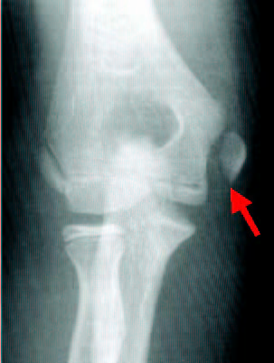
In younger athletes, their growth plates are the weak link and more prone to an injury. Growth plate injuries instead of ligament injury are more common in those athletes. Medial epicondylar avulsion fractures can happen instead of UCL tear. The sublime tubercle, where UCL inserts on ulna, can also avulse. In addition to valgus stress to the medial part of the elbow, the lateral side of the joint is under compressive force. This force can also cause an injury to the lateral side such as OCD (osteochondritis dessecans).
Throwing mechanics plays an important role in reducing such stress to the joint. Certain mechanics are known to put more stress. Thus, it becomes important that overhead athletes are taught the right mechanics at the beginning because it is a lot harder to re-learn the right way after learning faulty motion than to learn the correct mechanics from the beginning.
Treatment of these conditions include cessation of any overhead activities including throwing, physical therapy to strengthen rotator cuff muscles especially posterior cuff and forearm musculature if it is pain-free, ice, NSAID's to reduce inflammation in an acute phase, etc. Then, gradual interval throwing program should be initiated. If conservative treatment fails, surgical treatment will be necessary.
Mechanisms of injury involves valgus stress and traction force from the pronator-flexor mass complex. During baseball pitching, the ligament may be under stress that is close to the maximum torque that it can take. A repetitive stress to the ligament over a period of time may cause a partial or complete tear of the ligament.
In younger athletes, their growth plates are the weak link and more prone to an injury. Growth plate injuries instead of ligament injury are more common in those athletes. Medial epicondylar avulsion fractures can happen instead of UCL tear. The sublime tubercle, where UCL inserts on ulna, can also avulse. In addition to valgus stress to the medial part of the elbow, the lateral side of the joint is under compressive force. This force can also cause an injury to the lateral side such as OCD (osteochondritis dessecans).
Throwing mechanics plays an important role in reducing such stress to the joint. Certain mechanics are known to put more stress. Thus, it becomes important that overhead athletes are taught the right mechanics at the beginning because it is a lot harder to re-learn the right way after learning faulty motion than to learn the correct mechanics from the beginning.
Treatment of these conditions include cessation of any overhead activities including throwing, physical therapy to strengthen rotator cuff muscles especially posterior cuff and forearm musculature if it is pain-free, ice, NSAID's to reduce inflammation in an acute phase, etc. Then, gradual interval throwing program should be initiated. If conservative treatment fails, surgical treatment will be necessary.
![]() |
| MRI image of torn UCL |
![]() |
| X-ray of medial epicondylar avulsion fracture |
Monday, September 13, 2010
ACL In Preadolescents: Should It Be Reconstructed??
Even though it is still rare, ACL tear in preadolescent athletes are more talked about and diagnosed. A lot more ACL tears in adolescents occur than in preadolescents. However, it is a huge problem to athletes with torn ACL. Whether or not to reconstruct the torn ACL in still controversial.
ACL connects two bone in the knee, femur and tibila, thigh bone and shine bone. It provides stability to the knee and keeps the shine bone from sliding forward on femur and it also prevents excess rotational movements of the knee. Without ACL, athletes will not be likely to perform athletic maneuvers 100% even though there are some who do.
In the preadolescent, their growth plates (physes) are open and still growing. Some doctors are not willing to operate in those athletes. They usually wait until a growth spur occurs to reconstruct the torn ACL. Because there are some reports that growth arrest or other complications may happen if they put a hole through growth plates. There are also reports that say that it is ok to put a hole through growth plates unless a hardware is left in the middle of them.
There are a few methods to reconstruct ACL without drilling a hope in growth plates. One is to put a graft between epiphyes. Each end of the graft will not reach the growth plate and stopped before. Another way is to take an IT band and use is as a graft. Many orthopedic surgeons will wait until growth spur is finished to reconstruct the torn ACL. One problem that may occur is that leaving a torn ACL in the knee will likely lead to degenerative damage in the knee, thus, leading to premature OA (osteoarthritis). Think about 20 some year old female suffering from OA?! That is not easy.
In the end, there is no perfect answer to this question, I believe. There are pros and cons either way, early reconstruction or delayed reconstruction. They all should be discussed by the doctor and family member including the injured athlete herself/himself.
ACL connects two bone in the knee, femur and tibila, thigh bone and shine bone. It provides stability to the knee and keeps the shine bone from sliding forward on femur and it also prevents excess rotational movements of the knee. Without ACL, athletes will not be likely to perform athletic maneuvers 100% even though there are some who do.
In the preadolescent, their growth plates (physes) are open and still growing. Some doctors are not willing to operate in those athletes. They usually wait until a growth spur occurs to reconstruct the torn ACL. Because there are some reports that growth arrest or other complications may happen if they put a hole through growth plates. There are also reports that say that it is ok to put a hole through growth plates unless a hardware is left in the middle of them.
There are a few methods to reconstruct ACL without drilling a hope in growth plates. One is to put a graft between epiphyes. Each end of the graft will not reach the growth plate and stopped before. Another way is to take an IT band and use is as a graft. Many orthopedic surgeons will wait until growth spur is finished to reconstruct the torn ACL. One problem that may occur is that leaving a torn ACL in the knee will likely lead to degenerative damage in the knee, thus, leading to premature OA (osteoarthritis). Think about 20 some year old female suffering from OA?! That is not easy.
In the end, there is no perfect answer to this question, I believe. There are pros and cons either way, early reconstruction or delayed reconstruction. They all should be discussed by the doctor and family member including the injured athlete herself/himself.
Subscribe to:
Posts (Atom)